The Scan That Remembered: Tan Mu’s MRI and the Brain That Held Its Breath
In 2019, during a deep freediving session, Tan Mu experienced cerebral hypoxia. The condition is what it sounds like: the brain is deprived of oxygen, and the deprivation affects the hippocampus first, because the hippocampus is the structure where memories are formed and retrieved, and it is among the most metabolically demanding regions of the brain, consuming roughly twenty percent of the body's oxygen despite accounting for only two percent of its mass. When the oxygen supply drops, the hippocampus stops functioning before the other structures do, and the result is a gap, a period of time during which the brain was operating but not recording, during which the person was alive and awake and doing things but unable to form the memories that would allow her to remember doing them later. Tan Mu lost time. She surfaced from the dive and could not recall what had happened during the period when her hippocampus was not functioning, and the loss was not abstract. It was specific. It was a gap in the record, a section of the magnetic tape erased, a stretch of the hard drive written over with zeroes, a period of her life that she had lived but could not access because the organ that stores access had been temporarily disabled by the very activity that had required it to function. She went to the hospital. She had an MRI scan. The scan showed her brain from the inside, a cross-sectional image in which the gray matter and the white matter and the cerebrospinal fluid and the ventricles were rendered in shades of gray and black that corresponded to their tissue density, and the image showed a brain that was intact, which meant that the hypoxia had not caused structural damage, which meant that the memory loss was functional rather than anatomical, which meant that the organ was fine but the recording had failed, which meant that the problem was not in the hardware but in the process, not in the substance of the brain but in its operation, not in what the brain was but in what it was doing, and the distinction between what the brain is and what it does is the distinction that the painting makes.
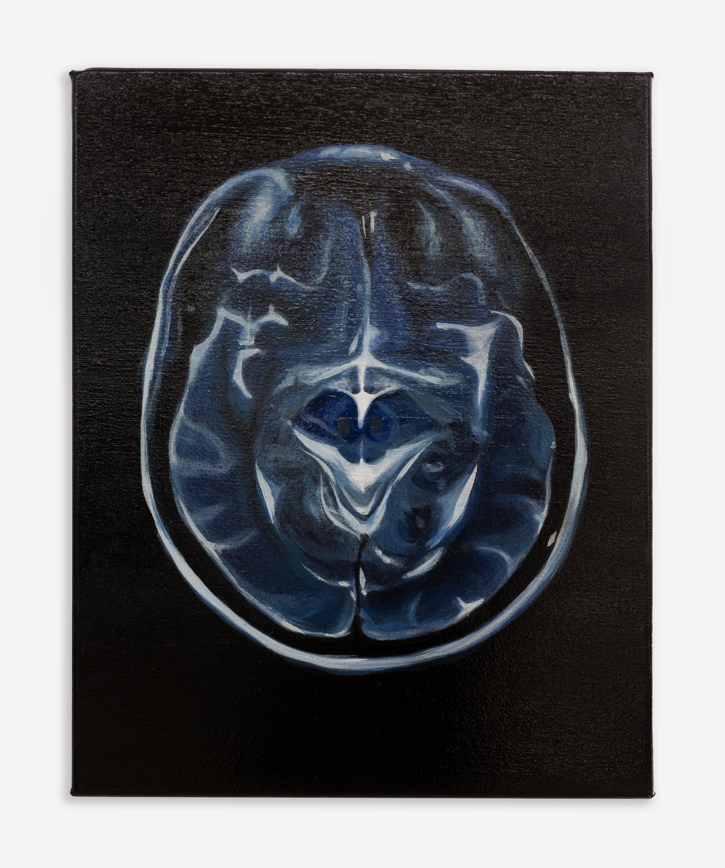
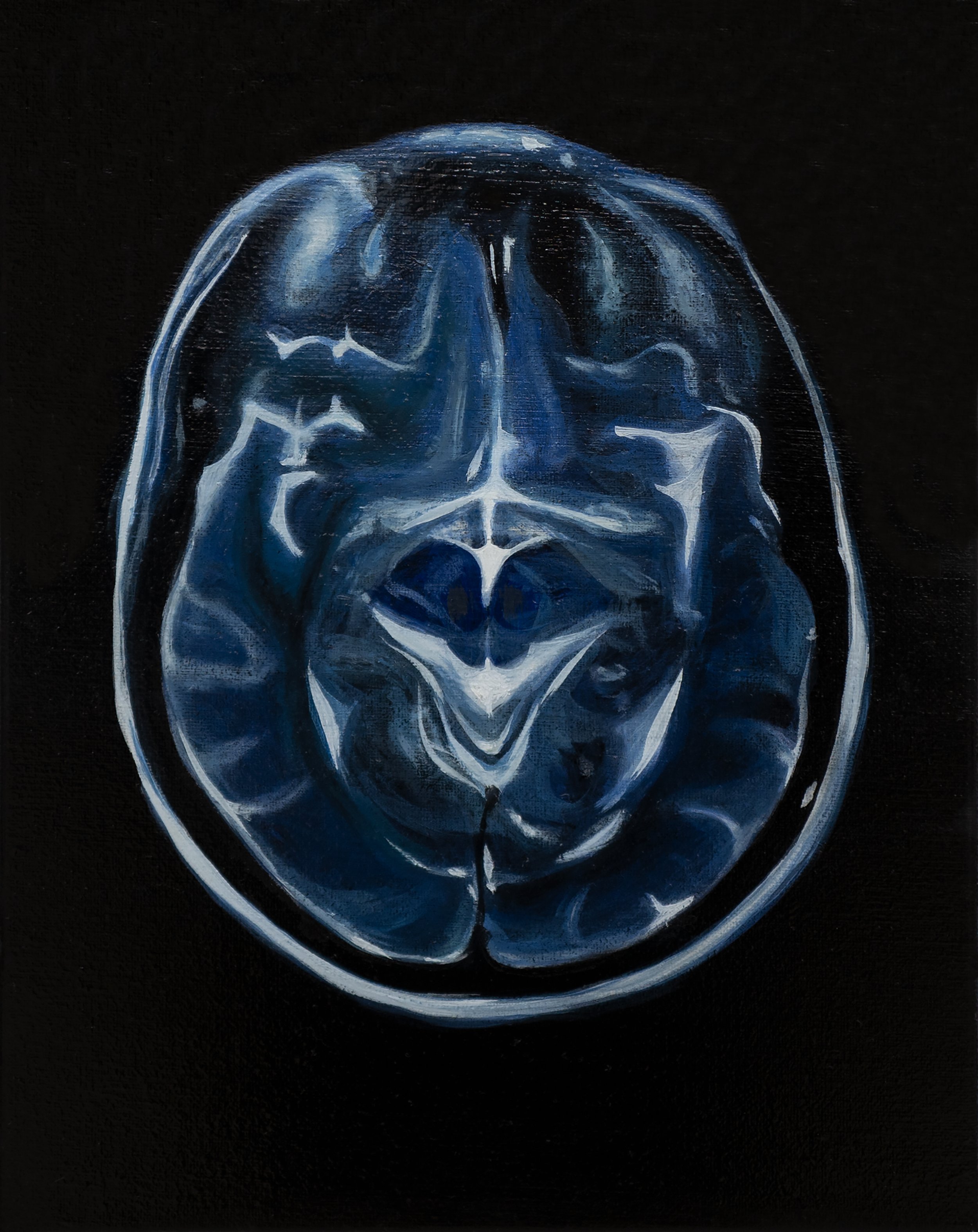
MRI is a small painting, 36 by 28 centimeters, oil on linen, roughly the size of a sheet of A4 paper, roughly the size of a brain scan as it appears on a medical monitor or a printed film. The scale is intimate and diagnostic, the scale of a clinical image that a doctor holds in her hand while discussing the results with the patient, the scale of the information that a person receives about the organ that is producing the reception of the information. The composition is a cross-sectional view of a brain, the kind of image that magnetic resonance imaging produces, a horizontal slice through the organ that reveals the two hemispheres of the cerebrum, the folded cortex of the gray matter, the branching white matter beneath it, the dark spaces of the ventricles, and the surrounding cerebrospinal fluid that appears as a bright border between the brain and the skull. The palette is restricted to the colors of the MRI itself: grays ranging from near-white to near-black, with touches of warm amber in the regions of highest metabolic activity where the blood flow is densest, and cool blues in the cerebrospinal fluid that surrounds the brain and fills the ventricles. These colors are not arbitrary. They correspond to the actual appearance of a T2-weighted MRI scan, in which the fluid appears bright and the dense tissue appears dark, inverting the visual expectation that the solid matter would be the most visible element, so that the brain in the painting reads as a form surrounded by a luminous field rather than a form that contains the field within it.
The paint handling enacts the distinction between structure and function that the subject demands. The gray matter, the cortex with its ridges and valleys, is painted with precision, each sulcus and gyrus defined by a thin line of darker gray against a lighter gray ground, the way a high-resolution MRI resolves the folds of the brain with a clarity that allows the radiologist to identify individual structures. The white matter beneath the cortex is rendered more diffusely, the boundaries between one region and another softening as the paint moves inward from the surface, the brushwork becoming broader and less defined, so that the interior of the brain reads as a space where clarity diminishes, where the resolution drops, where the scanner's ability to distinguish one tissue from another decreases and the image becomes a gradient rather than a map. The ventricles, the fluid-filled cavities at the center of the brain, are the brightest elements in the painting, rendered in a pale blue-gray that catches the light, because on a T2-weighted scan the fluid is the brightest signal, and the brightness is not an aesthetic choice but a translation of the physics of magnetic resonance, the way hydrogen protons in water molecules respond to the radio frequency pulse that the machine sends through the patient's body, the way the protons emit a signal that the machine receives and translates into an image in which the water is bright and the tissue is dark and the brain appears as a structure that is mostly made of water, mostly made of fluid, mostly made of the same substance that fills the ocean and falls from the sky and circulates through every organ of every body on the planet.
Francis Bacon painted heads throughout his career, but the heads that matter for this argument are the ones from the early 1950s, specifically the series of papal portraits that he began after seeing Diego Velazquez's Portrait of Pope Innocent X in reproduction and that culminated in Study after Velazquez's Portrait of Pope Innocent X, completed in 1953 and now in the collection of the Des Moines Art Center. The painting depicts a figure seated on a throne, enclosed in a vertical frame of horizontal lines that suggest both a glass box and a surgical curtain, the kind of partition that divides a hospital room into compartments for examination. The figure's face is distorted, the mouth open in a scream that may or may not be producing sound, the eyes half-visible behind smears of paint that suggest both the blur of a photograph taken in motion and the disintegration of a face that is being erased by the act of looking at it. The body beneath the face is barely present, a shadow of purple and black that suggests mass without specifying it, a silhouette that occupies the space of a body without revealing the body's details, the way an X-ray reveals the shape of the skeleton without revealing the flesh that surrounds it. Bacon was not painting a pope. He was painting a subject under observation, a figure who had been placed in a box and examined, a body that was being looked at by a machine that produced an image of the body's interior, and the scream was the body's response to being seen in this way, the protest of the organism against the gaze that reduces it to data.
The parallel to Tan Mu's MRI is structural rather than visual. Bacon and Tan Mu are both painting the body as it appears to a technology that sees through it, that renders the surface transparent and the interior visible, that produces an image in which the body is not a person but a pattern of densities, a map of tissues that can be read by a specialist who does not need to know the person whose body produced the image. The MRI scan and the Bacon painting share this condition: they both depict a body that is being observed by a technology, and the technology's observation is not neutral. It produces an image that transforms the body from a subject into an object, from a person who is looking into a person who is being looked at, from a consciousness that is experiencing to a specimen that is being examined. The transformation is the subject of both works. Bacon's figure screams because the examination has made the body into a spectacle. Tan Mu's brain scan is the product of the same examination, rendered not with horror but with the precision of a painter who has undergone the examination and emerged from it with an image that she chose to translate into paint, not as a protest against the medical gaze but as an acceptance of it, an acknowledgment that the gaze has seen something real, that the interior of the body is not a private space that the technology has invaded but a structure that the technology has revealed, and that the revelation, however uncomfortable, however reductive, however different from the experience of being the person whose brain is being scanned, is also a form of knowledge that the person did not have before the scan and that the person cannot now forget, because the image is in her memory now, alongside the gap that the hypoxia created, the gap that the scan was meant to investigate, the gap that the scan could not fill because the scan shows structure and the gap is a failure of function, and structure and function are not the same thing even though they occur in the same organ at the same time.
The hippocampus is a small structure, roughly the shape and size of a seahorse, located in the medial temporal lobe of each hemisphere. It is the brain's memory engine, the place where short-term memories are consolidated into long-term memories, the place where the experiences of the day are replayed during sleep and transferred from temporary storage to permanent storage, the place where the brain decides what to keep and what to discard. When the hippocampus is deprived of oxygen, it stops forming memories. The person continues to act, to speak, to respond, because the other structures of the brain, the cortex, the basal ganglia, the cerebellum, the brainstem, are still receiving enough oxygen to maintain basic function, but the recording mechanism has been suspended, and the result is a period of consciousness without memory, a period during which the person is present but the person's hippocampus is not, a period that will later appear as a blank, a gap, a stretch of time that was lived but not recorded, an experience that happened but cannot be retrieved because the organ that stores retrievals was temporarily offline. Tan Mu has described this experience directly: after the hypoxia, she experienced gaps in memory that made her acutely aware of both the complexity and the fragility of the brain as a container for memory. The painting that resulted, MRI, is not a painting of the hypoxia itself. It is a painting of the scan that was performed to determine whether the hypoxia had caused structural damage, and the scan showed that it had not, which meant that the memory loss was functional, which meant that the hippocampus would recover, which meant that the gap was temporary, but the gap had already occurred, and the experience of the gap could not be recovered because the gap was precisely the period during which the recording mechanism was not operating, so the experience of the gap is known to Tan Mu only by its absence, only by the fact that there is a period in her life that she cannot remember, a blank in the record that the record itself cannot explain because the record is the thing that was blank.
The book that Tan Mu read during this period, The Three Pound Universe by Judith Hooper and Dick Teresi, gave her a framework for understanding what had happened. The title refers to the approximate weight of the adult human brain, three pounds of tissue that contains roughly 86 billion neurons, each connected to thousands of other neurons through synapses, producing a network of roughly 100 trillion connections, a number so large that it exceeds the number of stars in the Milky Way galaxy and approaches the number of stars in the observable universe. The book's argument, which resonated with Tan Mu's experience, is that the brain is a three-pound universe, a microcosm that mirrors the macrocosm, a structure whose organization recapitulates the organization of systems at other scales, from the branching of neural dendrites that resembles the branching of river systems, to the distribution of synaptic connections that resembles the distribution of galaxies in the cosmic web, to the way that information flows through the brain from one region to another that resembles the way that energy flows through ecosystems from one trophic level to the next. The comparison is not metaphorical in the loose sense. It is structural. The brain, the river system, and the cosmic web all exhibit the same kind of organization: a small number of hubs with a large number of connections, a large number of nodes with a small number of connections, and a pattern of flow that follows the path of least resistance from one hub to another, producing a network that is efficient, resilient, and vulnerable to the same kinds of failure, the failure of a single hub that disconnects large regions of the network from the rest, the failure of a single oxygen supply that disables the hippocampus and erases the record of everything that happened while it was offline.
Gerhard Richter's 48 Portraits, completed in 1972 and now in the collection of the Museum of Modern Art in New York, consists of forty-eight paintings of forty-eight men, each derived from a black-and-white photographic source, each rendered in oil on canvas at a scale slightly smaller than life size, each depicting the head and shoulders of a figure against a neutral gray background. The series was originally commissioned for the German Pavilion at the 1972 Venice Biennale and was intended to represent a pantheon of modern Western intellectuals, scientists, writers, and philosophers whose images Richter sourced from encyclopedia entries and photographic archives. The paintings are recognizably derived from photographs. The brushwork is flat, the details are soft, the focus is slightly blurred, the way a photograph is slightly blurred when it is reproduced at a scale larger than its resolution can support, and this blurriness is not an accident of technique but a deliberate effect, Richter's method of translating the photographic image into paint while preserving the photographic quality of the original, the quality that distinguishes a photograph from a painting, which is the quality of having been made by a machine rather than by a hand, the quality of objectivity, the quality of the mechanical eye that records what is in front of it without selecting or emphasizing or interpreting, the quality that makes the photograph a document rather than an artwork, the quality that Richter wanted to preserve even as he translated the document into the medium of painting, which is the medium of selection and emphasis and interpretation, the medium of the hand.
The connection to Tan Mu's MRI is precise. Both works take images produced by machines, images that were generated by technologies of seeing that extend the human eye beyond the visible spectrum, and translate them into paint. Richter's source images were produced by photographic cameras that captured reflected light. Tan Mu's source image was produced by a magnetic resonance imaging machine that captured the radio-frequency emissions of hydrogen protons in the brain's water molecules, a technology that does not see in the way that a camera sees but that constructs an image from data, an image that corresponds to the physical properties of the tissue rather than to its visual appearance, an image that shows the brain not as it looks to the eye, which cannot see inside the skull, but as it looks to the machine, which can see through the skull by measuring the behavior of the protons that make up the brain's water content. The MRI image is a construction. It is a mathematical reconstruction of the proton signals, a map of tissue densities rendered in grayscale, a representation of the brain's interior that is accurate to the structure but alien to the experience of being the person whose brain is being represented. Tan Mu's translation preserves this alienation. The painting does not try to make the brain look familiar. It does not anthropomorphize the scan. It translates the scan's grayscale palette into paint, its resolution into brushwork, its clinical distance into the intimacy of a small canvas held in the hand, and the translation produces a work in which the viewer can see both the image and the technology that produced it, both the brain and the machine that rendered it visible, both the person whose brain was scanned and the gap that the scan was meant to investigate, the gap that the scan could not fill because the gap was not structural but functional, not in the tissue but in the operation, not in what the brain was but in what it failed to do during the minutes when the oxygen was gone and the hippocampus stopped recording and the person kept living but the record went blank and the only evidence that the gap existed was the gap itself, the absence of memory, the silence in the archive, the three-pound universe holding its breath.