The Clone and the Canvas: Tan Mu's Epithelial Cells and the Color of Lineage
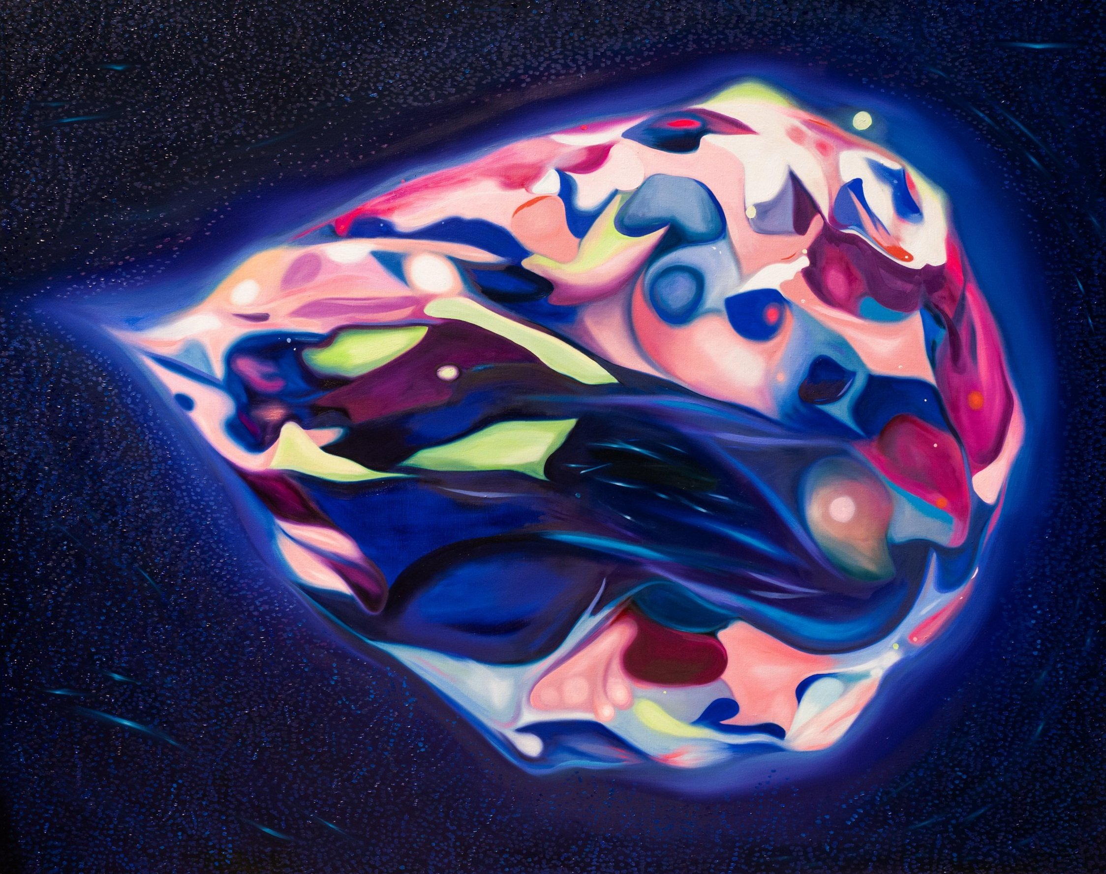
At thirty centimeters from the surface, the painting is a geology of pigment. Clusters of saturated color, emerald and ruby and sapphire and topaz, sit raised against a dark ground like mineral deposits in a vein of rock. Each cluster has edges where the paint thickens into a ridge before dropping away to the linen, and the linen itself is visible in the gaps between clusters, a pale weave that reads as the substrate beneath a layer of tissue. At this distance, you see the hand's work: the way a single cluster contains three or four distinct shades of the same hue, each one a separate decision, a separate application of the brush, each one tracing the boundary of a cell group that shares a single ancestor. The dots that fill the background, smaller than a pinhead, resolve at this range into individual marks, each one placed with enough deliberation that you can see the point where the brush lifted and the paint pulled to a tiny peak. You are close enough to count the cells in a single cluster. You are close enough to see that the color is not flat but layered, that the emerald cluster contains a vein of lighter green where the paint was applied more thinly, that the ruby cluster has a shadow of darker red where a second coat was laid over the first. At this distance, the painting is not a representation of cells. It is a construction of them.
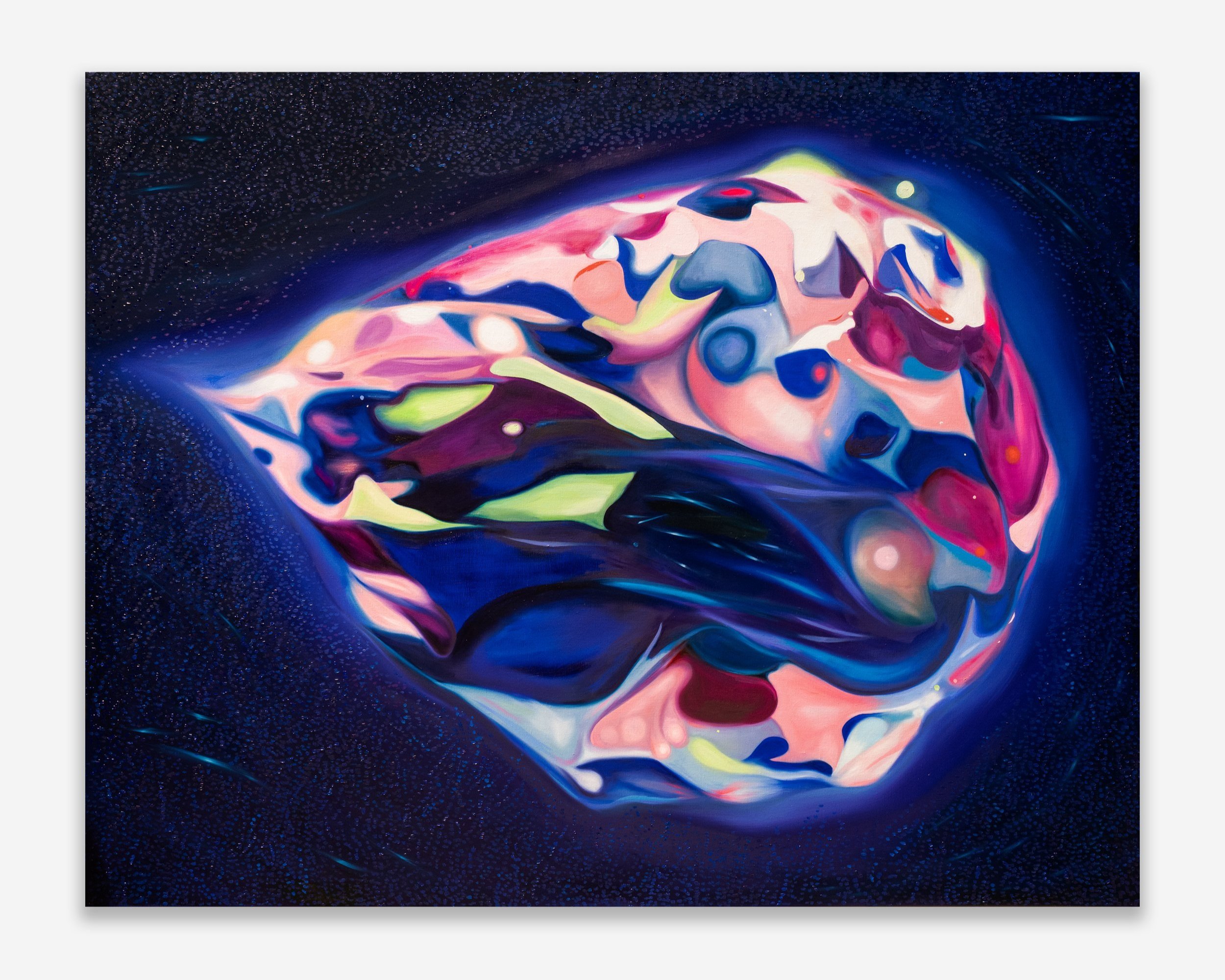
Step back two meters and the construction reassembles into a field. The clusters now read as territories, each one a colored province in a map of cellular migration. The gaps between them become the borders of a cellular nation state. The background dots, which at close range were individual marks, now read as a population, a scattering of particles that could be biological debris, could be data points, could be the dust of a culture medium in which something is growing. The painting holds both readings simultaneously: the cellular and the cartographic, the biological and the informational. This is the double vision that clonal labeling produces. In the laboratory, researchers stain individual cells with fluorescent proteins that mark their lineage. Every daughter cell inherits the color of its parent. A single green cell divides, and its descendants form a green territory. A single red cell divides, and its descendants form a red territory. The color is not decorative. It is a record of descent. It tells you who came from whom. It makes lineage visible as pigment, which is to say, it makes lineage visible in the same medium that Tan Mu uses to paint it.
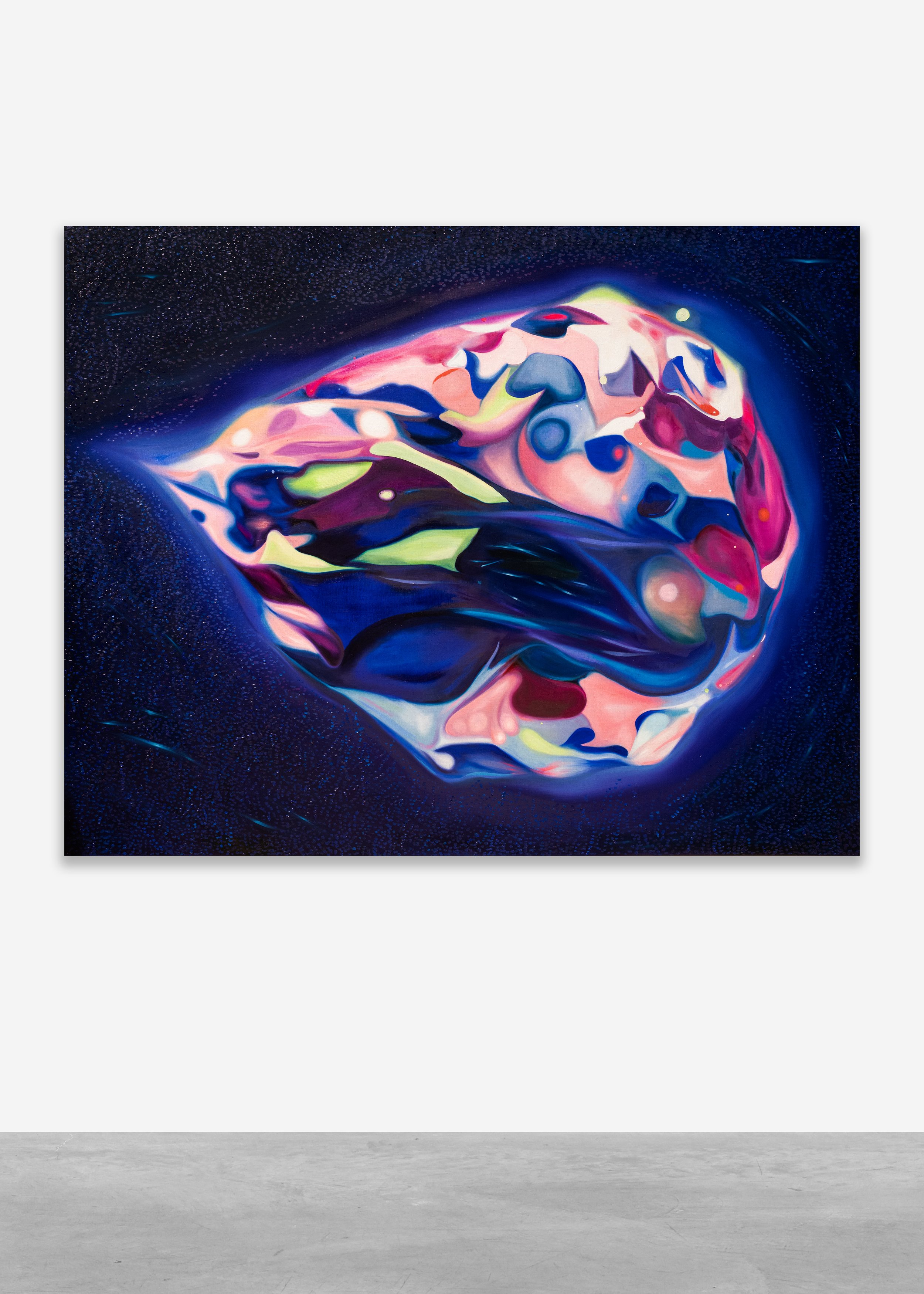
Epithelial Cells (2024) is a painting about human epithelial cells growing in a dish. The cells have been clonally labeled, meaning that cells of the same color are descendants of a single cell at the time of labeling. This scientific approach helps quantify cell proliferation and reveals how proliferative potential is passed from stem cells to their differentiated daughter cells. The painting is 122 x 153 cm (48 x 60 in), oil on linen, a format large enough that the clusters of color spread across the canvas like archipelagos seen from a satellite. The medium is listed simply as oil on linen, but the paint behaves differently in different regions of the canvas. In the cellular clusters, it sits thick and waxy, built up in layers that catch raking light and create actual topographic variation on the surface. In the background, the dots are applied more thinly, sitting closer to the linen, as if the cells themselves were growing on top of a substrate of information. The contrast in paint application is not accidental. It mirrors the biological reality: the cells are the substance, and the culture medium, the substrate, the nutrient solution in which they float, is the medium that sustains them. The paint makes this distinction physical. The clusters protrude. The background recedes. The painting has a topography, and the topography indexes a hierarchy of biological significance.
The colors themselves are not natural in the sense that they reproduce the actual appearance of stained cells under a microscope. They are natural in a different sense: they are the colors of gemstones. Tan Mu has described her longstanding attraction to gemstones and mineral structures, their saturated colors and internal complexity. When she encountered laboratory images of epithelial cells stained for scientific analysis, she recognized in them the same visual richness she found in geological specimens. The emerald of a beryl crystal. The ruby of corundum. The sapphire of a Kashmir stone. Each cluster in the painting glows with the internal luminosity of a gem, and this luminosity is not merely aesthetic. It is the luminosity of information encoded as color, of data made visible through staining protocols that are, in their own way, as precise and as interpretive as the mixing of pigment on a palette. The parallel between scientific staining and painterly color mixing is one that Tan Mu has noted explicitly: "In my own work, variations within a single color emerge through pigment mixtures that share the same origin but differ in proportion." The same red, mixed with more white or more yellow or more medium, produces a family of reds that are related by origin but differentiated by ratio. This is precisely how clonal labeling works. The same genetic lineage, expressed through slightly different conditions of growth and differentiation, produces a family of cells that are related by descent but differentiated by function. The painting does not merely depict this logic. It enacts it.
Ernst Haeckel's Kunstformen der Natur (Art Forms of Nature), published in installments between 1899 and 1904 and collected as a single volume in 1904, is one of the most sustained attempts in the history of science to make the argument that biological structure is aesthetic structure. Haeckel, a German zoologist and evolutionary biologist, produced over a hundred lithographic plates depicting radiolarians, medusae, bryozoa, and other organisms from the deep sea and the microscopic world. His drawings are not neutral illustrations. They are compositions. Each plate arranges its subjects in patterns that emphasize symmetry, repetition, and radial order. The radiolarians, single-celled organisms whose mineral skeletons take the form of intricate geodesic domes, are rendered with the precision of an architectural draftsman and the compositional instinct of a painter. Haeckel believed that the aesthetic properties of these organisms were not incidental to their biology. He coined the term "ecology" in 1866, and he understood the relationships between organisms and their environments as having an aesthetic dimension. The beauty of the radiolarian skeleton was not separate from its function. The structure that allowed the organism to float in the water column was the same structure that produced the geometric regularity that Haeckel found so compelling.
The structural parallel with Tan Mu's Epithelial Cells is not merely visual, though the visual resonance is immediate. Both works present biological structures as compositions of color and form that exceed the requirements of scientific illustration. Both insist that what the microscope reveals is not ugly or neutral or purely functional but is, in its own register, beautiful. Both translate the microscopic into the visible through an act of selection, emphasis, and composition that is as much aesthetic as it is documentary. But the parallel runs deeper than shared admiration for biological form. Haeckel's project was taxonomic. He drew these organisms in order to classify them, to place them in an evolutionary tree that would demonstrate the unity of all life. His symmetry was an argument about descent. The regularity of the radiolarian skeleton was, for Haeckel, evidence of the regularity of natural law. Tan Mu's project is also taxonomic, but her taxonomy is not evolutionary. It is clonal. The clusters of color in Epithelial Cells are not arranged to demonstrate the unity of life across species. They are arranged to demonstrate the unity of descent within a single tissue. Each color is a lineage. Each cluster is a family tree compressed into a two-dimensional map. Where Haeckel used symmetry to argue that all life shares a common origin, Tan Mu uses color to argue that all the cells in a single tissue carry the memory of a specific origin. The painting is not a tree of life. It is a family portrait of a single layer of skin.
Epithelial cells are the body's border guards. They form tightly packed layers that line every surface where the interior meets the exterior: the skin, the gut, the airway, the blood vessel. They are the cells that negotiate exchange and protection, that decide what comes in and what stays out. Tan Mu chose them for this reason. "These cells exist at boundaries," she writes in her Q&A, "constantly negotiating exchange and protection." The choice connects to Julien Offray de La Mettrie's L'Homme Machine (Man a Machine, 1748), a text that Tan Mu cites directly. La Mettrie argued that the human body is a complex, highly organized machine, and that consciousness, sensation, and volition are products of its mechanical organization rather than evidence of an immaterial soul. The epithelial cell is a machine in La Mettrie's sense. It opens and closes junctions, admits and blocks molecules, proliferates and differentiates according to programs encoded in its DNA. It operates without will, without intention, without the interiority that we associate with conscious beings. And yet it is, in Tan Mu's painting, luminous, structured, and organized with a precision that evokes not mechanism but artistry.
The clonal labeling technique that the painting depicts is itself a form of machine vision. Researchers introduce fluorescent proteins into individual cells using genetic engineering. The proteins are passed to daughter cells during division. Under the right illumination, each lineage glows with its own color. The technique allows scientists to track the fate of individual stem cells over time, to see which lineages expand and which contract, which differentiate and which remain undifferentiated. It is a way of making the invisible visible, of rendering the passage of descent as a trace of color. The technique was developed in the early 2000s and has become central to cancer research, because the behavior of clonal populations in a tissue culture dish mirrors the behavior of tumor cells in a body. A clone that expands too aggressively, that outcompetes its neighbors for space and nutrients, is a tumor in miniature. The painting, by depicting these clonal territories at a scale that a viewer can walk toward and walk away from, makes the relationship between normal growth and pathological growth legible. The territory that glows emerald is not inherently benign. The territory that glows ruby is not inherently malignant. The color tells you who the cells came from. It does not tell you what they will do next.
Tan Mu places this work in explicit dialogue with her earlier Chromosomes (2022), which also reflects on genetic structure and cellular information. But where Chromosomes presents the genome as a static architecture, a blueprint laid out for inspection, Epithelial Cells presents the cell as a dynamic entity, a living process that unfolds in time. The clonal clusters in the painting are not still. They are spreading. They are competing. They are growing toward the edges of the dish, which is to say the edges of the canvas, which is to say the boundaries of the painting itself. The composition suggests a moment of expansion frozen mid-process, a snapshot of biological activity that will look different an hour from now, a day from now. This temporal quality distinguishes the painting from scientific illustration, which typically aims for a representative moment that could serve as a generalized reference. Tan Mu's painting does not generalize. It particularizes. It shows you a specific dish, a specific distribution of clonal populations, a specific ratio of emerald to ruby to sapphire to topaz. The specificity is the point. The painting is not about epithelial cells in general. It is about these epithelial cells, in this dish, at this moment in their proliferation.
Louise Bourgeois's Cell series, produced between 1989 and 1993, is an unlikely but structurally revealing parallel. Bourgeois constructed enclosed spaces, each one a room-sized installation containing objects that carried personal and symbolic resonance: a guillotine, a child's dress, a bulb, a chair. The cells were architectural, but they were also biological. Bourgeois spoke of them as representations of the psyche, and the title itself, Cell, invokes the basic unit of biological life. Each cell was a bounded space with an interior that could not be fully accessed from the exterior. The viewer could look in through gaps in the enclosure, but could not enter. The interior remained partially hidden, partially legible, always withholding more than it revealed.
The parallel with Tan Mu's Epithelial Cells operates at the level of the boundary. Bourgeois's cells are enclosures that define an interior against an exterior. The epithelial cells that Tan Mu paints are biological enclosures that perform the same function at microscopic scale. Both artists are preoccupied with the membrane: the structure that separates one domain from another, that maintains identity by regulating what passes through. For Bourgeois, the membrane is architectural, a door or a wall or a cage. For Tan Mu, the membrane is cellular, a lipid bilayer that opens and closes ion channels and admits nutrients and expels waste. But in both cases, the membrane is not merely a barrier. It is a site of negotiation. It determines what the interior becomes by determining what the interior receives. The epithelial cell's identity, its decision to differentiate into a protective barrier cell or a secretory cell or a sensory cell, is shaped by what crosses its membrane. Bourgeois's installations, similarly, gain their meaning from what they contain and what they exclude. The objects inside the cell are not random. They are selected, curated, placed. They constitute an identity that the enclosure both protects and presents. The cell, in both artists' hands, is not a neutral container. It is a portrait of its contents, and its contents are a portrait of the forces that shaped them.
The difference is one of scale and access. Bourgeois's cells are human-scale. You walk around them. You peer through their doors. Your body is roughly the same size as the enclosure, and the relationship between your body and the cell is one of parity, of mutual sizing. Tan Mu's cells are microscopic, painted at a scale that makes a single cell the size of a coin. The relationship between the viewer's body and the painted cells is one of radical disproportion. You stand before a field of entities that are, in life, one hundredth of a millimeter across, and they are rendered large enough that you can see the variation in their color, the thickness of their boundaries, the density of their populations. This disproportion is not a distortion. It is a translation. The painting takes something that is below the threshold of unaided vision and brings it into the register of the seen. It performs the same function as the microscope, but it performs it through paint rather than optics, and in doing so it introduces something that the microscope cannot: the hand, the palette, the decision about which color to emphasize, which boundary to sharpen, which cluster to let dissolve into the field. The painting is a stained preparation, but the staining is done by a painter, not a biologist, and the choices embedded in the staining carry an argument that no laboratory protocol could produce.
Saul Appelbaum, writing in his 2025 essay "Dreaming in Public," describes Tan Mu's paintings as operating through a process of "arbitration," a term he borrows from electro-acoustic music systems: an input passes through a process of decision and emerges as an output. The concept applies to Epithelial Cells with particular force. The laboratory image is the input. The painting is the output. The arbitration occurs in the space between, in the decisions that Tan Mu makes about color, scale, emphasis, and surface. These decisions are not arbitrary. They are informed by years of looking at gemstones, at mineral structures, at the way light passes through translucent materials and is absorbed by opaque ones. They are informed by her training at the Fine Arts School Affiliated to China Central Academy of Fine Arts in Beijing, where the philosophical principle of ge wu zhi zhi, investigating things to extend knowledge, shaped her approach to research-driven painting. They are informed by her sustained engagement with the microscopic world, from MRI (2021) to Synapse (2023) to Chromosomes (2022) to this work. Each painting in this sequence is an arbitration of a different biological image, and each arbitration produces a different output that carries the trace of the decision process that shaped it. The gem-like colors in Epithelial Cells are the trace of a decision to emphasize the mineral quality of cellular structure, to draw out the parallel between the crystal and the colony, between the geological and the biological. The raised surface of the clusters is the trace of a decision to make the cells physically present, to give them a topographic reality that a flat application of paint would not possess. The dot background is the trace of a decision to place the cells in a field of information, to suggest that what surrounds the cell is not empty space but a medium dense with data.
The ethical dimension that Tan Mu identifies in her Q&A is not separate from the visual argument. "As genetic decoding and editing technologies advance, we are forced to confront difficult issues," she writes. "How far should human intervention go. Who has access to these technologies. Could their misuse deepen global inequalities or redefine concepts of life itself." These questions are present in the painting not as annotations but as properties of the image itself. The clonal labeling technique that the painting depicts is a precursor to gene editing. The same fluorescent proteins that mark lineage can be attached to the CRISPR-Cas9 system that edits genes. The step from marking a cell's origin to modifying a cell's destiny is shorter than it appears. The painting, by depicting the moment before the edit, the moment when lineage is visible but unmodified, holds open the question of what comes next. The clusters are growing. The populations are expanding. The territories are shifting. What the painting shows is a tissue at a point of possibility, a moment when the future of each lineage has not yet been determined by intervention. The beauty of the image, its gem-like luminosity, its compositional balance, its chromatic richness, is the beauty of a moment that will not last. In the laboratory, the cells in the dish will continue to divide. They will reach the edges of the culture medium. They will exhaust their nutrients. They will die, or they will be harvested, or they will be genetically modified to serve a purpose that the original cell could not have foreseen. The painting preserves a moment that the biology will not. This is what painting does that the microscope cannot. It holds time still. It allows you to stand at the edge of the dish and look at the cells before they become something else, before the emerald lineage is edited to express a therapeutic protein, before the ruby lineage is deleted because it carries a mutation, before the entire population is replaced by a clone that someone designed in a laboratory and grew in a dish and painted on a canvas and called Epithelial Cells.
The painting's background of scattered dots, Tan Mu's signature technique, operates across her practice as a field of information that connects the microscopic to the cosmic. In Epithelial Cells, the dots can be read as biological particles, data points, or celestial bodies, and this ambiguity is deliberate. "These dots can be read as biological particles, data points, or even celestial bodies," Tan Mu writes, "suggesting that information flows across scales from the microscopic to the cosmic." The dot field is not background in the passive sense. It is the connective tissue of the painting, the medium through which meaning moves from one register to another. The epithelial cell is a unit of biological information. The data point is a unit of digital information. The celestial body is a unit of cosmological information. All three are points of light against a dark field. All three are instances of structure emerging from what would otherwise be formless. The painting holds them in the same visual space, and in doing so it argues that the distinction between biological, digital, and cosmological scales is a distinction of degree, not of kind. The cell and the star and the data point are all structures that organize information, and they are all visible in the same way: as luminous points in a dark field. The painting does not resolve the ambiguity. It maintains it. It allows the dots to be all three things simultaneously, and it asks the viewer to hold that multiplicity, to see the cell and the star and the data point in the same mark, to understand that the painting's argument is not about epithelial cells alone but about the structural logic that epithelial cells share with every other form of organized information in the universe.
What remains when the painting has made all of these arguments is the surface itself. The thick clusters of waxy oil paint that protrude from the linen. The thin dots that sit flush against the weave. The contrast between the raised and the flat, the substantial and the ephemeral, the cellular and the informational. The painting is a tissue, not in the metaphorical sense but in the material sense: a layer of substance on a substrate, a skin of paint on a body of linen. The epithelial cells that Tan Mu paints are themselves tissues in this material sense. They grow on a substrate. They form a layer. They have a topography. The painting does not illustrate this structure. It shares it. The paint is the cell, and the linen is the dish, and the dots are the medium, and the light that falls on the surface and catches the ridges of the clusters is the same light that falls on a culture dish in a laboratory and makes the fluorescent proteins glow. The painting does not represent the glow. It produces a glow of its own, through a different mechanism, at a different scale, for a different purpose. The purpose is to make you see what the biologist sees when she looks through the microscope, and to make you feel what she feels when she sees it: that the interior of the body is as organized, as structured, as beautiful, and as dangerous as the exterior of the world. The boundary that the epithelial cell maintains is the boundary between what we are and what we might become. The painting sits on that boundary, and it glows.